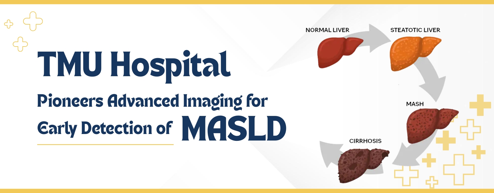
Teerthanker Mahaveer University Hospital, Moradabad, Uttar Pradesh, is at the forefront of clinical innovation with the adoption of advanced imaging techniques for the precise evaluation of Metabolic Dysfunction-Associated Steatotic Liver Disease (MASLD), a condition previously known as NAFLD or NASH.
In a detailed academic insight authored by Prof. (Dr.) Rajul Rastogi, Department of Radiodiagnosis at Teerthanker Mahaveer Medical College & Research Centre, has comprehensively addressed the growing significance of imaging in diagnosing MASLD. The article highlights the limitations of conventional ultrasound and the superiority of new modalities like Ultrasound-Derived Fat Fraction (UDFF) and Proton-Derived Fat Fraction (PDFF) MRI, both actively employed at TMU Hospital.
MASLD arises primarily from metabolic conditions such as type 2 diabetes mellitus, obesity, and hyperlipidaemia, marking a shift from the traditional causes of liver cirrhosis, like chronic alcoholism and viral hepatitis. As cirrhosis remains irreversible without liver transplantation, early identification and management of steatosis are pivotal.
While ultrasonography (USG) is widely used to detect fatty liver, it is largely qualitative and subjective. The diagnosis often hinges on increased liver echogenicity relative to the kidneys. This approach can be misleading in conditions like chronic viral hepatitis or congestive hepatomegaly, which also present with elevated echogenicity.
Furthermore, the grading of fatty liver into Grades I, II, and III lacks precision and cannot reliably track minor changes during treatment or disease progression.
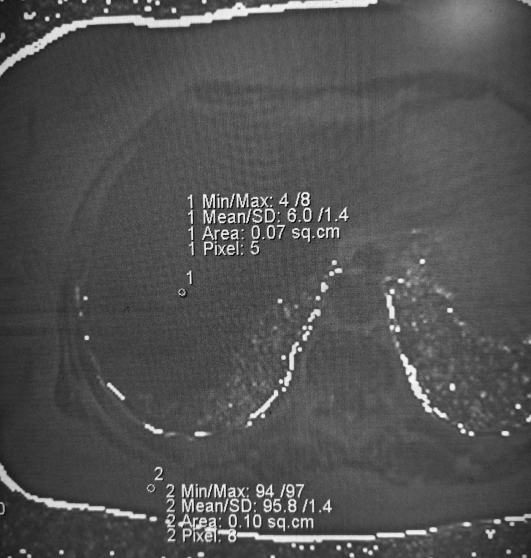
The Ultrasound-Derived Fat Fraction (UDFF) technique, now available at TMU Hospital, represents a significant upgrade over routine USG. It quantifies liver fat content objectively, allowing clinicians to distinguish between true fatty liver and echogenic appearances due to other conditions.
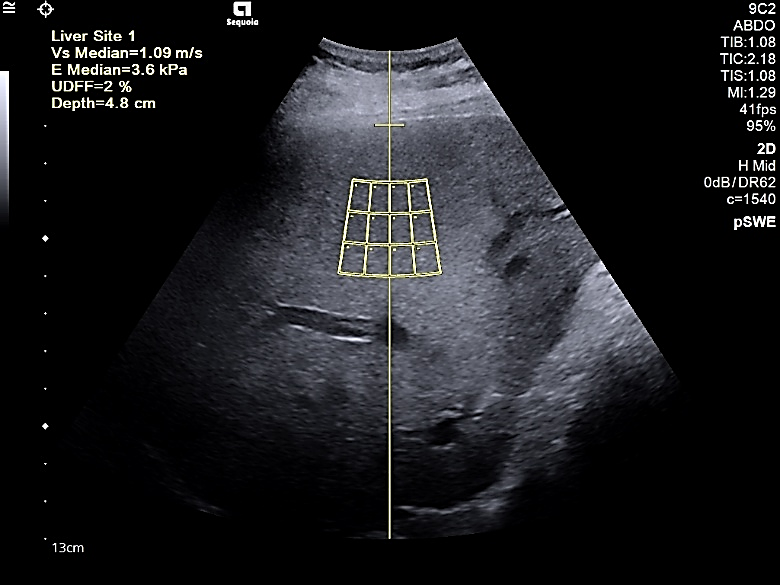
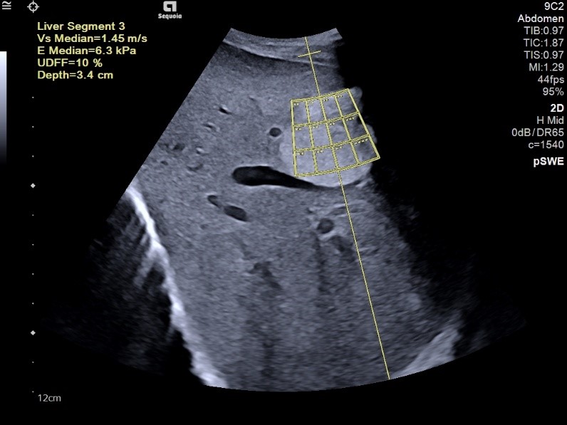
These capabilities reduce unnecessary biopsies and cross-sectional imaging, streamlining diagnosis and enhancing patient safety.
While MRI-based PDFF remains the gold standard for hepatic fat quantification, its high cost, longer scan times, and specialised software requirements limit widespread use. Nevertheless, TMU Hospital continues to provide this advanced imaging technique for critical cases requiring high accuracy.
With these imaging advancements, TMU Hospital offers a robust framework for early MASLD detection, enabling timely lifestyle and therapeutic interventions. This proactive approach significantly curbs the progression to cirrhosis, reducing the long-term burden on healthcare systems.
Dr. Rajul Rastogi emphasises that integrating UDFF and PDFF into routine liver assessments has transformed diagnostic radiology's role in managing metabolic liver diseases.
MD (Radiodiagnosis), PG Dip MSK USG (Spain)
 Role of Digital Mammography in Detection...
© TMU Hospital is Proudly Owned by TMU